
Figure 21.
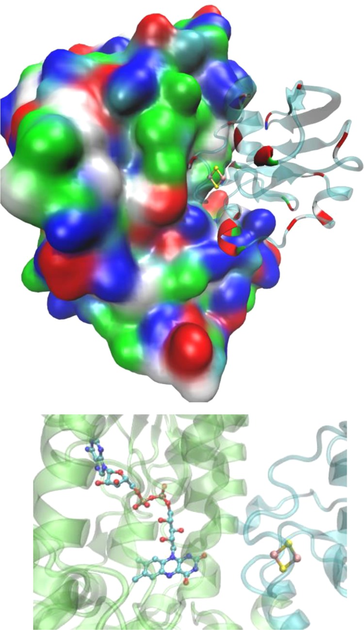
Structure of adrenodoxin (right) in complex with adrenodoxin reductase (left) (PDB ID 1E6E). As shown, red acidic patches of adrenodoxin are positioned against blue basic residues of adrenodoxin reductase. A zoom-in region of the cofactors (Fe–S and FAD) is shown at the bottom.