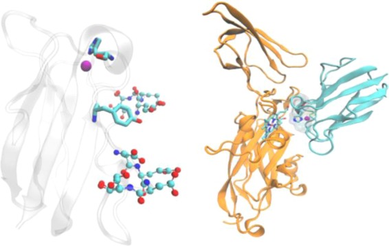
Figure 51.

Structures of plastocyanin (left) and the complex of plastocyanin and cyt f (right). Left: copper ion is represented as a purple ball, His87 and Tyr 83 are represented in licorice format, and residues in two acidic patches are represented as ball and stick models. Right: plastocyanin is colored cyan, and cyt f is orange. The copper ion and His87 from plastocyanin and heme from cyt f are also shown.
