
Figure 69.
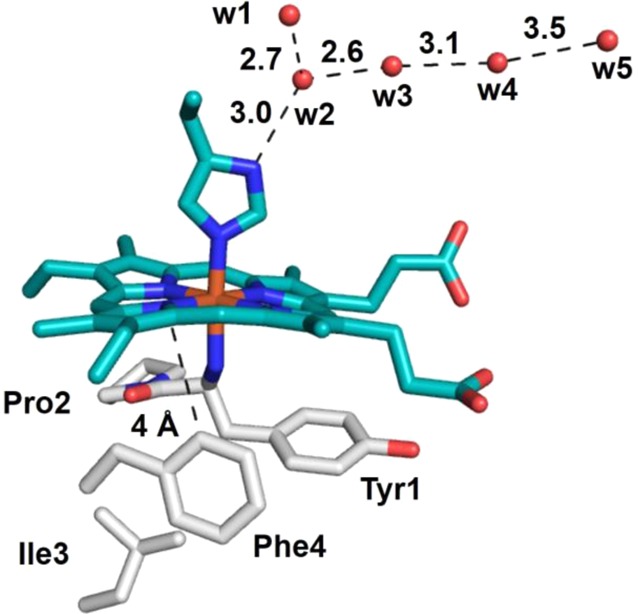
Environment around the heme of cyt f (PDB ID 1HCZ). Hydrophobic residues are shown as gray sticks. The “edge-to-face” interaction at 4 Å between Phe4 and the heme that is proposed to be important to tune the reduction potential of the heme iron is shown. The five conserved molecules that have been proposed to act as “proton wires” that couple ET with proton transfer are shown as red spheres. Residue numbering of waters is arbitrary.