Figure 6.
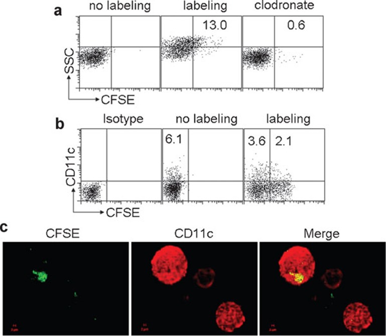
Generation of MPs containing Lm components by macrophages and uptake by DC in vivo. (a) MPs were mainly generated by macrophages after peritoneal infection of Lm. BALB/c mice (n=6) with or without 48 h macrophage depletion by clodrolip were i.p. injected with 1×10⁷ CFU CFSE-labeled Lm. Twelve hours later, MPs were isolated from the peritoneal lavage fluids and analyzed by flow cytometry. (b, c) MPs were taken up by DCs in vivo. The isolated CFSE-MPs after 1×10⁷ CFU CFSE-labeled Lm peritoneal infection were i.p. injected into naive BALB/c mice. Six hours later, the peritoneal cells were harvested and stained with CD11c mAb and analyzed by flow cytometry (b) or observed under two-photon laser scanning fluorescent microscope (c). CFSE, carboxyfluorescein succinimidyl ester; CFU, colony forming unit; DC, dendritic cell; i.p., intraperitoneally; Lm, Listeria monocytogenes; mAb, monoclonal antibody; MP, microparticle.

