Figure 2.
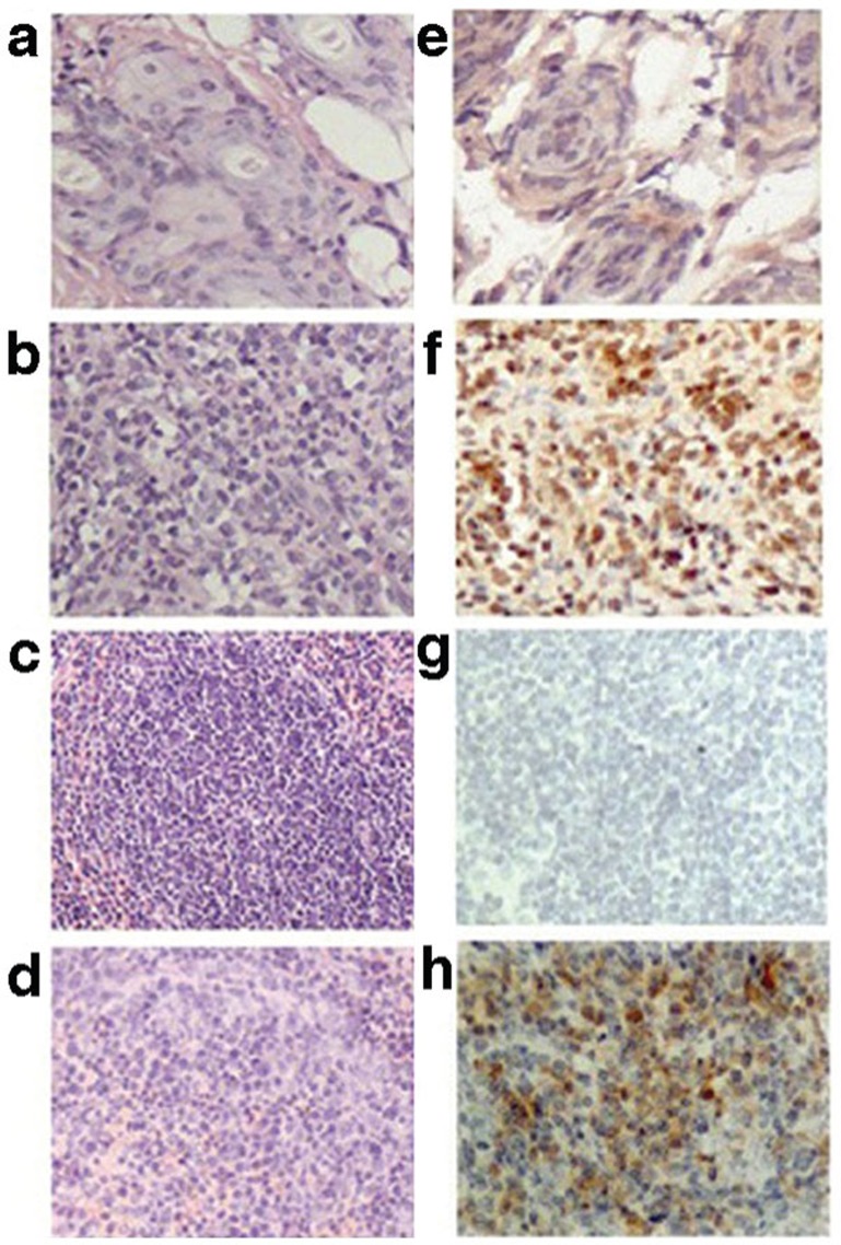
Expression of Pim2 within grafted skin and spleen. The left panels show sections with hematoxylin and eosinstaining, and the right panels show immunohistochemical staining using anti-Pim2. (a, e) Isograft skin. (b, f) Allograft skin. (c, g) Splenic areas of isografted mice. (d, h) Splenic areas of allografted mice. The results have been repeated in three independent experiments.

