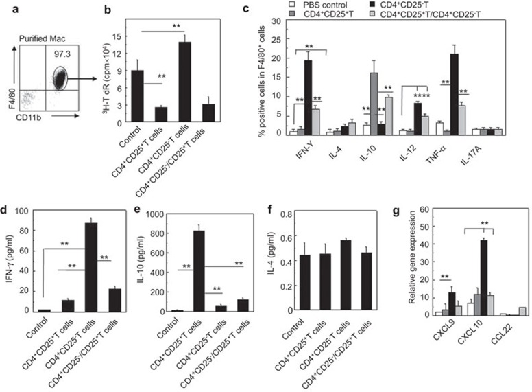
Figure 3.
Altered immunogenicity and cytokine and chemokine secretion of recipient macrophages induced by allogeneic donor CD4+CD25+ Tregs in NOD-scid mice. (a) The recipient resident macrophages were isolated and the purity was confirmed with CD11b and F4/80 double staining by FCM. (b) The immunogenicity of the recipient resident macrophages was assessed with MLR. The proliferation of BALB/c CD4+ T cells induced by the allogeneic macrophages isolated from NOD-scid mice that received no cells, CD4+CD25+ T cells, CD4+CD25− T cells or both CD4+CD25+ T cells and CD4+CD25− T cells was determined by 3H-TdR incorporation in vitro. (c) Cytokine production by recipient F4/80+ macrophages was determined by two-color intracellular staining FCM. The percentages of IFN-γ+, IL-4+, IL-10+, IL-12+, TNF-α+ and IL-17A+ cells in F4/80+ macrophages were determined after the cells were stimulated with LPS (100 ng/ml). (d–f) The cytokine levels in the peripheral blood of recipient NOD-scid mice were assessed by enzyme-linked immunosorbent assayA 3 days after the adoptive transfer of the indicated cells. (g) The relative mRNA expression of the chemokines, CXCL9, CXCL10 and CCL22, in the peritoneal macrophages of the recipient NOD-scid mice, was analyzed by real-time PCR. More than five mice in each group were assayed. The data are the mean±s.d. **P<0.01 among the indicated groups. FCM, flow cytometry; IFN, interferon; LPS, lipopolysaccharide; MLR, mixed lymphocyte reaction, TNF, tumor-necrosis factor; Treg, regulatory T cell.

