
Figure 1.
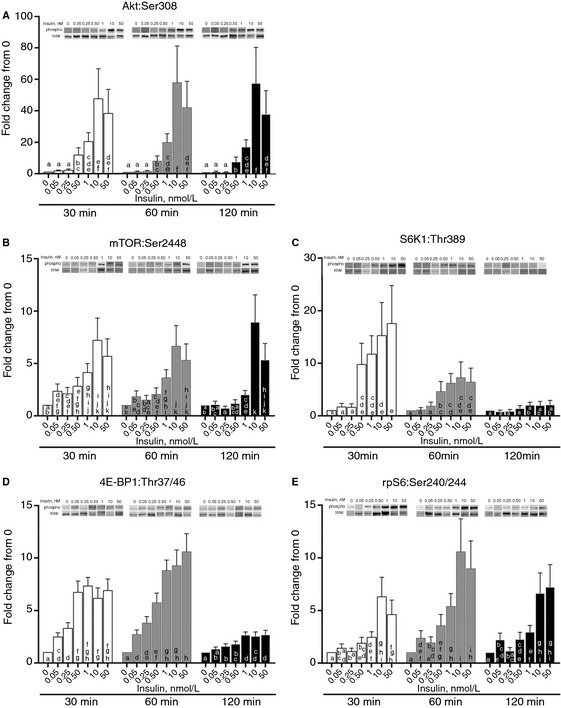
The effect of insulin concentrations on Akt and mTOR signaling. Different (0.05, 0.25, 0.5, 1, 10, and 50 nmol/L) concentrations of insulin (in HBS) were incubated with myotubes for 30, 60, and 120 min. Myotubes were lysed and protein extracts were analyzed using western blotting. (A) Phosphorylation status of AktSer308. abcdefColumns with uncommon letters differ, P = 0.9921. (B) Phosphorylation status of mTORS er2448. abcdefghijkColumns with uncommon letters differ, P = 0.3123. (C) Phosphorylation status of S6K1Thr389. abcdeColumns with uncommon letters differ, P = 0.0376. (D) Phosphorylation status of 4E‐BP1Thr37/46. abcdefghColumns with uncommon letters differ, P < 0.0001. (E) Phosphorylation status of ribosomal protein S6Ser240/244. abcdefghiColumns with uncommon letters differ, P = 0.775. Resulting images are displayed from a representative experiment above each graph. For arrangement of samples in gels for electrophoresis, all time points for two samples were run on a single gel. Thus, all samples were not run on a single gel/blot. Data are mean ± SEM and are presented as phospho/total made relative to baseline.