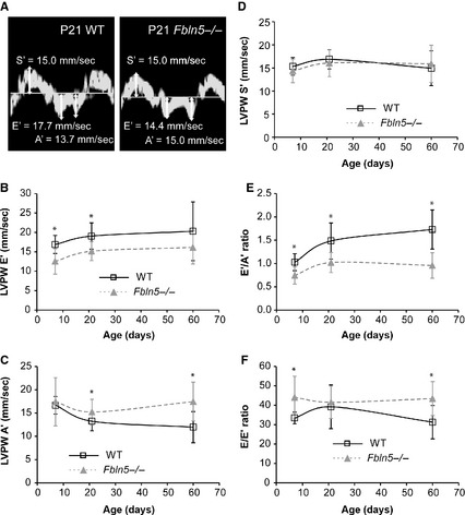
Figure 8.

Representative tissue Doppler images (A) with left ventricular (LV) posterior wall (PW) velocities during systole (S’) and during early (E’) and atrial (A’) filling in P21 wild‐type (WT) and Fbln5−/− mice. LVPW E’ (B) velocity is decreased in Fbln5−/− mice at P7 and 21. LVPW A’ (C) velocity is increased in Fbln5−/− mice at P21 and P60. LVPW S’ (D) velocity is similar between genotypes at all ages. The E’/A’ (E) and E/E’ (F) ratios, which are indices of diastolic function, are different in Fbln5−/− mice compared to WT at most ages. *P < 0.05 between genotypes. n = 10–16 per group.
