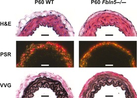
Figure 10.

Representative histology sections of P60 carotid arteries. H&E staining and picrosirius red staining (PSR) observed under polarized light show no obvious differences between genotypes. Verhoeff Van Gieson (VVG) staining shows less intense staining of the elastic lamellae in Fbln5−/− arteries, especially in the outer layers. Scale bars = 30 μm. Histology sections from six different mice were examined for each group.
