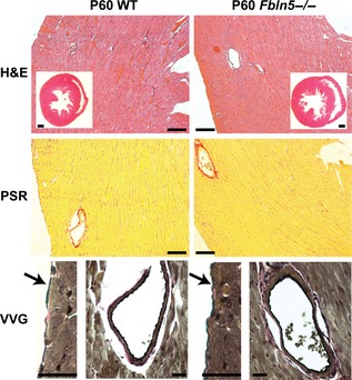
Figure 11.

Representative histology sections of P60 mouse hearts. H&E staining of the entire heart and of the left ventricular (LV) free wall show no differences between genotypes. Picrosirius red (PSR) staining of the LV free wall shows no differences in collagen density between genotypes. Verhoeff Van Gieson (VVG) staining of the thin pericardial layer (arrow) shows intact elastic fibers in both genotypes. VVG staining of coronary vessels shows an intact internal elastic lamina in both genotypes and no elastin staining between cardiomyocyte layers. H&E inset scale bars = 500 μm, H&E and PSR scale bars = 100 μm, VVG scale bars = 20 μm. Histology sections from six different mice were examined for each group.
