
Figure 2. RNA foci and C9RAN protein pathology are present in various regions of the central nervous system in C9FTLD/ALS.
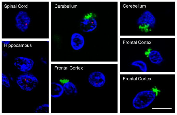
Fluorescence in situ hybridization of C9FTLD/ALS tissue sections using a probe against sense G4C2 transcripts (spinal cord, hippocampus) or antisense C4G2 transcripts (cerebellum, frontal cortex) was followed by immunofluorescence staining to detect poly(GP) inclusions. Note that RNA foci (red) in the nucleus (stained with DAPI, blue), and star-shaped cytoplasmic poly-GP inclusions (green), seldom co-occur in the same cells. Scale bar = 10 μm.