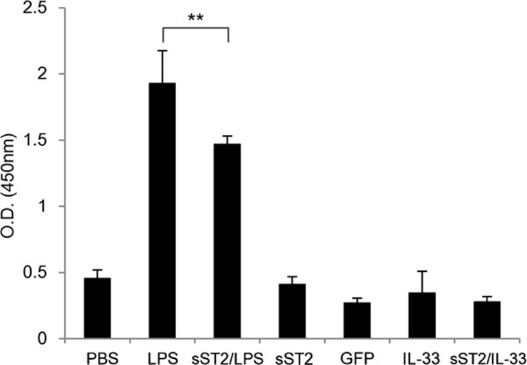
Figure 4.
sST2 attenuates the proliferation of naive T-lymph cells by stimulated MDDCs autologously. iMDDCs were seeded at 2×10⁴/well in 96-well tissue culture plates and cocultured with naive CD4+ T cells at a 1:10 ratio with or without 500 ng/ml LPS, 100 ng/ml IL-33, or 500 ng/ml GFP for 5 days. LPS-induced mMDDCs without T cells and PBS-treated iMDDCs were employed as controls. BrdU incorporation was measured to assay the proliferation of naive CD4+ T cells. The data are presented as the means of three experiments±s.d. A double asterisk denotes a statistically significant difference (**P<0.01). GFP, green fluorescent protein; IL, interleukin; iMDDCs, immature monocyte-derived dendritic cell; LPS, lipopolysaccharide; MDDC, monocyte-derived dendritic cell; mMDDC, mature monocyte-derived dendritic cell; PBS, phosphate-buffered saline; sST2, soluble ST2 protein.

