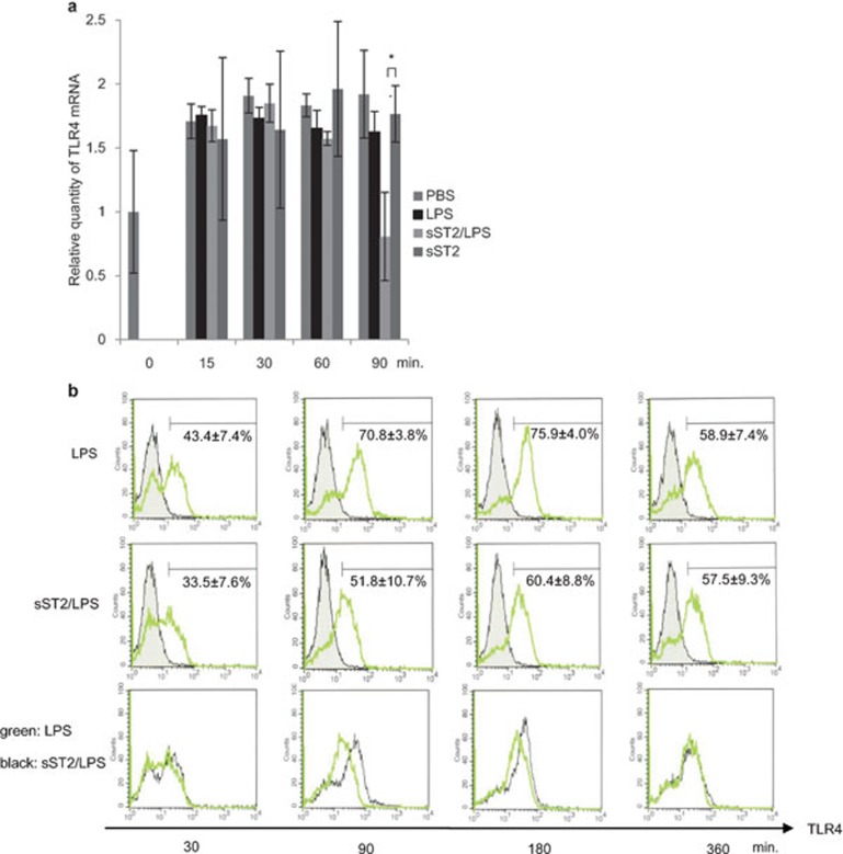
Figure 7.
TLR4 expression is suppressed by sST2. (a) Relative quantity of TLR4 mRNA was analyzed by real-time PCR after various stimuli (sST2, LPS, sST2/LPS) and normalized against endogenous β-actin. The data are presented as the means of three experiments±s.d. An asterisk denotes a statistically significant difference (*P<0.05). (b) iMDDCs were pre-treated with or without sST2 (100 ng/ml) and treated with LPS (500 ng/ml) for 30 to 360 min. TLR4 protein on the cell surface was analyzed by FACS with anti-TLR4-PE. The figures of the bottom column were showing the expression of TLR4 of LPS-stimulated iMDDCs (the upper column, red) superimposed on the middle column (sST2/LPS; green). Representative data of three experiments are presented. FACS, fluorescence-activated cell sorting; iMDDCs, immature monocyte-derived dendritic cell; LPS, lipopolysaccharide; sST2, soluble ST2 protein; TLR4, Toll-like receptor 4.

