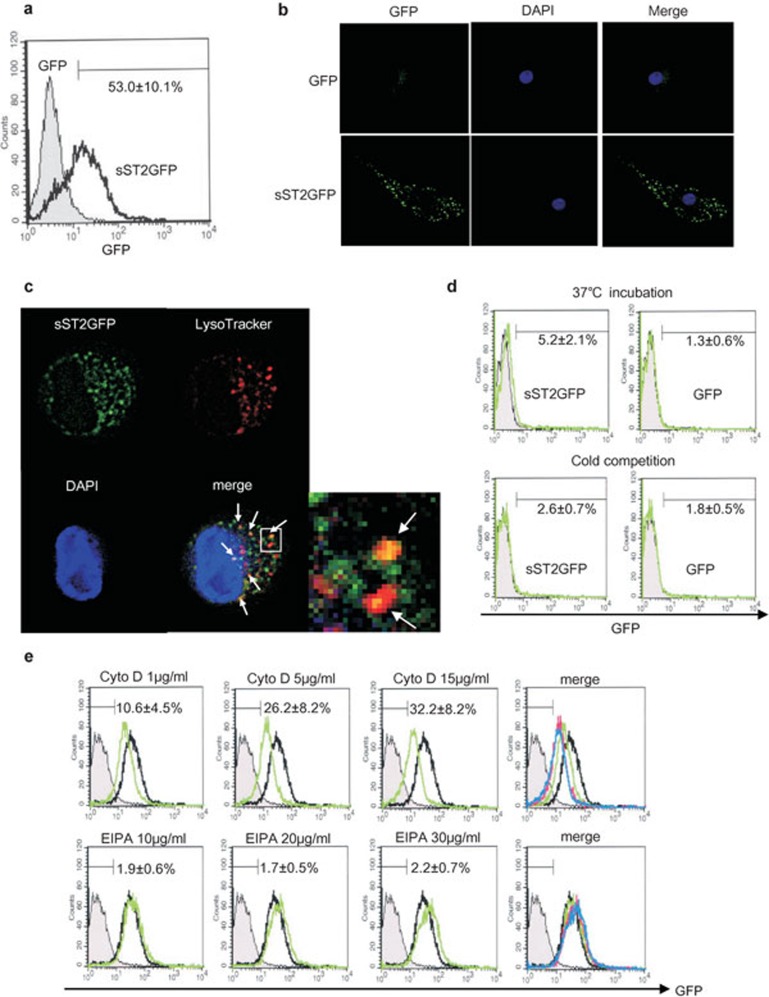
Figure 8.
sST2-GFP is internalized into iMDDCs. (a) iMDDCs were incubated with sST2-GFP (100 ng/ml) for 16 h at 37 °C. The fluorescence intensity of GFP was analyzed by FACS. One of five similar experiments is presented. The percentage of positive cells are indicated as the means of three experiments±s.d. (b) sST2-GFP is attached to iMDDCs which were incubated with sST2-GFP or GFP as described in (a) and observed with a fluorescence microscope (green, sST2-GFP; blue, DAPI). (c) sST2-GFP and lysosomes were co-localized in iMDDCs. The lysosomes in iMDDCs treated with sST2-GFP were stained with LysoTracker probes (Red DND-99) and observed with a fluorescence microscope (green, sST2-GFP; red, lysosome; yellow, sST2-GFP and the lysosome merged). The representative data of three similar experiments are presented. (d) Cold competition of sST2-GFP binding to iMDDCs. iMDDCs were incubated with sST2-GFP on ice or at 37 °C for 1 h. The fluorescence intensity of GFP was analyzed by FACS (upper panel green, 37 °C; lower panel green, on ice; gray area, unstained control). The percentage of positive cells are indicated as the means of five experiments±s.d. (e) Inhibition of macropinocytosis. iMDDCs were pre-treated with cytochalasin D (1, 5 and 15 µg/ml) or EIPA (10, 20 and 30 µg/ml) for 30 min, then sST2-GFP (100 ng/ml) was added for 16-h culture. After incubation, the cells were washed twice with PBS−, and then fluorescence was analyzed by FACS. The gray area shows the control without staining, and the black line shows iMDDCs incubated with sST2-GFP only. The merged panel presented cytochalasin D (green; 1 µg/ml, pink; 5 µg/ml, blue; 15 µg/ml) or EIPA (green; 10 µg/ml, pink; 20 µg/ml, blue; 30 µg/ml) lines. The percentage of positive cells are indicated as the means of five experiments±s.d. EIPA, 5-(N-ethyl-N-isopropyl)amirolide; DAPI, 4,6-diamidino-2-phenylindole; FACS, fluorescence-activated cell sorting; GFP, green fluorescent protein; iMDDCs, immature monocyte-derived dendritic cell; LPS, lipopolysaccharide; sST2, soluble ST2 protein.

