Figure 1.
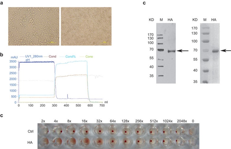
Expression of recombinant H5N1 HA proteins. (a) Representative images of Sf-9 cells before (left) and after (right) transfection of the recombinant baculovirus. (b) The elution curve of rHA proteins from an AKTA affinity chromatography system. (c) Purified rHA proteins were analyzed by western blot (left) with anti-HA antibodies and Coomassie blue staining of SDS–PAGE (right). M: protein marker. The arrows show the locations of the rHA proteins. (d) Hemagglutination activity analysis of the rHA proteins. HA, hemagglutinin; rHA, recombinant hemagglutinin.

