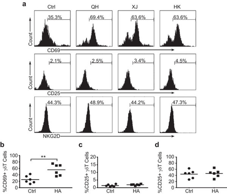
Figure 2.
Expression of early activation markers on γδ T cells in response to stimulation with rHAs from different H5N1 strains. (a) Flow cytometry analysis of the expression of the early activation markers CD69, CD25 and NKG2D on human peripheral γδ T cells in response to rHA stimulation. Representative images from at least three independent experiments. Human PBMCs were stimulated with recombinant QH-HA (A/Bar-headed Goose/Qinghai/2005 H5N1), XJ-HA (A/Xinjiang/2006 H5N1) or HK-HA (A/Hongkong/2003 H5N1). The cells were stained with antibodies specific for TCRγδ, CD69, CD25 and NKG2D. The TCRγδ-positive cells were gated, and the expression of CD69, CD25 or NKG2D was analyzed by flow cytometry. (b) The average percentage of CD69+γδ T cells from six independent experiments. (c) The average percentage of CD25+γδ T cells from six independent experiments. (d) The average percentage of NKG2D+γδ T cells from six independent experiments. **P<0.01. The horizontal lines represent the mean values. HA, hemagglutinin; PBMC, peripheral blood mononuclear cell; rHA, recombinant hemagglutinin.

