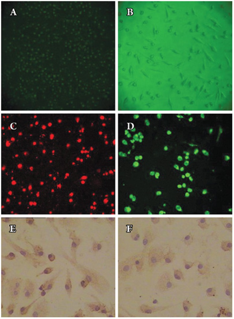
Figure 1.
Characterization of endothelial progenitor cell (EPC) number. (A) Mononuclear cells isolated from human peripheral blood appeared small and round under an inverted light microscope (original magnification×200). (B) After 7 days in culture, attached cells exhibited a spindle-shaped, endothelial cell-like morphology (original magnification×100). (C) DiLDL uptake (red, exciting wave-length 543 nm, original magnification×200) and (D) Adherent cells, lectin binding (green, exciting wave-length 477 nm, original magnification×200), were assessed under a laser scanning confocal microscope and by fluorescent microscopy. The cells were further analyzed by immunostaining with (E) vWF antibodies (original magnification×400) and (F) anti-CD31 (original magnification×400).

