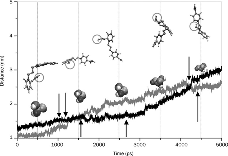
Figure 13.
Temporal spatial distance during SMD pulling between the center of mass of active-site Ser 203 and the N2 atom (grey line) and the N3 atom (black line) of the two pyridinium rings of HI-6 that was initially close to active triad and the peripheral site, respectively. Relative orientations and separations of the drug molecule with respect to Ser 203 (shown by space filling representation) are also shown at regular intervals. The grey circle encircling O1 atom of the drug is the oximate oxygen. See Supplementary information S-2 for the snapshots of protein-drug hydrogen bonds and water bridges at the time steps indicated in the figure by arrows.

