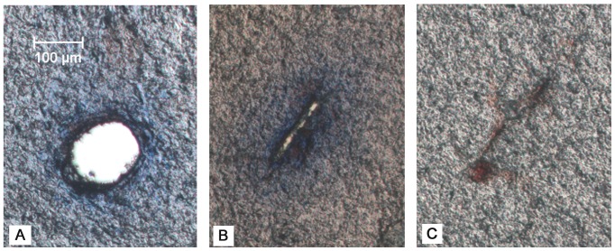
Figure 7. Different types of tissue damage left after needle retraction.
A) approximately circular hole surrounded by infused EBA; B) hole as a slender opening/crack; and C) red blood cell accumulation where it was difficult to measure the hole opening. All images are for fixed, 100 µm thick brain tissue slices.

