
Figure 11. Needle tissue damage observed with H&E staining.
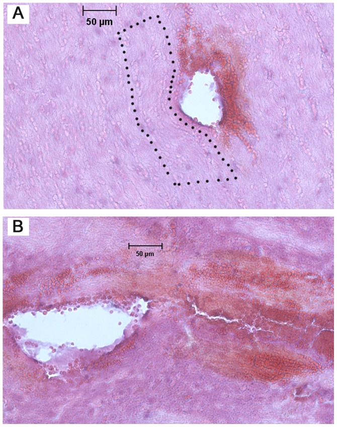
A) external capsule region showing deformation and compaction of surrounding tissue. In the enclosed area (dotted lines), layers of cells conform around the hole (needle insertion at 0.2 mm/s); B) CPu region showing extensive bleeding and tissue fracture at some distance away from of the hole (insertion at 10 mm/s).