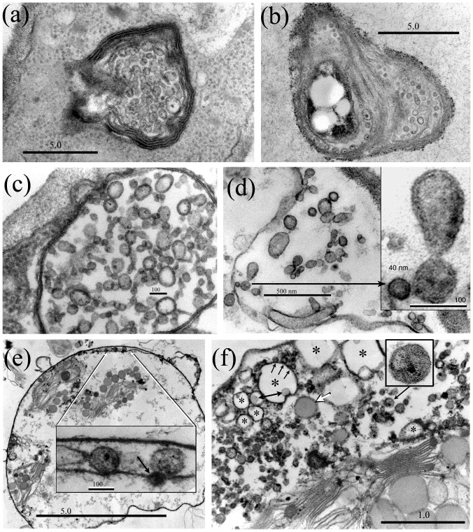
Figure 3. A. pullulans multivesicular bodies release budding Ms-vesicles.
(a) Ms-vesicles are formed within multivesicular bodies (MVB). (b) Similar vesicles are present within the periplasmic space of a dividing yeast cell (tangential section). (c) Mature MVB contain large numbers of budding vesicles, which are apparently released into the periplasmic space (d). (d) Inset: An enlarged budding vesicle. (e) Similar budding vesicles are observed within the envelope of modified Psilotum chloroplasts that may be enclosed by a fused fungal plasmalemma (note double-thickness of the outer membrane in the enlarged inset (e), and the ‘eruptions’ from the outer membrane). An opaque vesicle associated with the plastid inner membrane (inset e, arrow) may be budding into the plastid stroma. (f) An infected plastid containing large numbers of dividing vesicles within the expanded envelope. Most of the plastid inner membrane is missing: a short segment (visible right) is lined with vesicles, two with electron lucent centers (asterisk). The vesicles enlarge as electron dense bodies (white arrow), or as vacuole-like forms (asterisks). Note budding from the ‘vacuole’ margin (3 arrows) and presence of internal electron-dense bodies (double-ended arrow). The enlarged vesicle (3f) box) was cultured from modified Cuscuta subinclusa plastids. Bars = (a, b) 5.0 µm; (c) 100 nm; (d) 0.5 µm, inset 100 nm; (e) 5.0 µm, inset 100 nm; (f) 1.0 µm; boxed vesicle is 175 nm.

