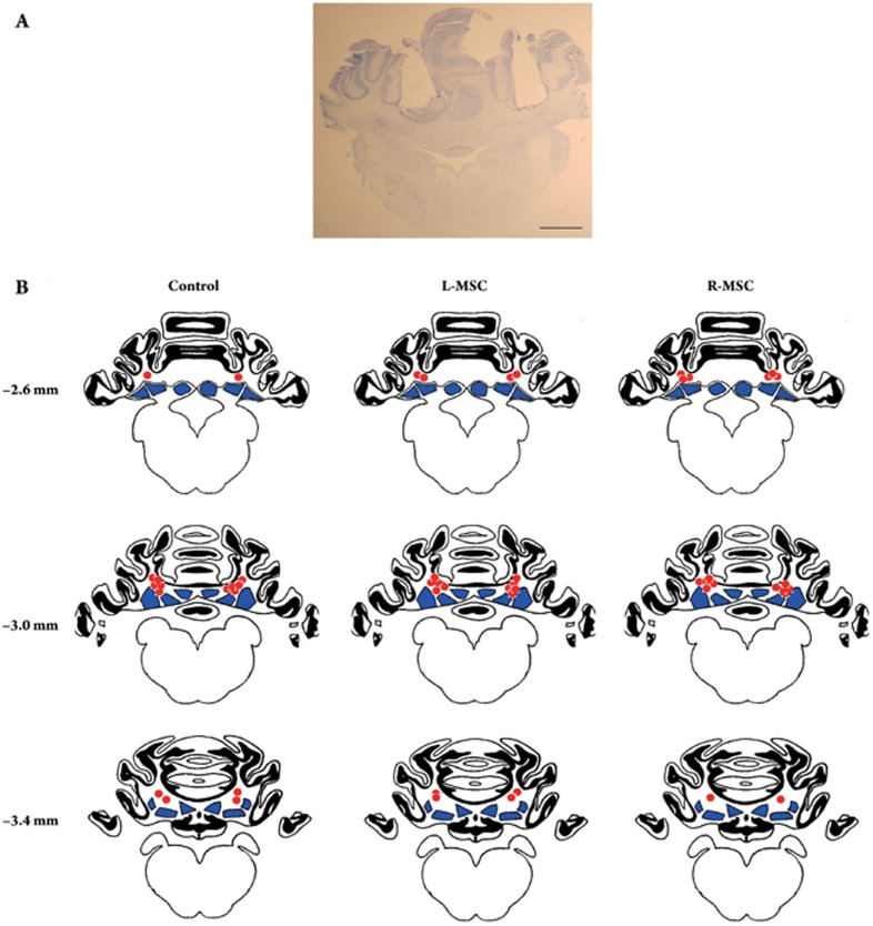
Figure 2.
Location of the drug microinjection sites in the cerebellum of guinea pigs. (A) Representative toluidine blue-stained coronal cerebellar section (10 μm) from a guinea pig that received drug microinjections. Two guiding cannulae pass through the ipsilateral and contralateral cerebellar cortex and their tips lay immediately dorsal to the deep cerebellar nuclei (DCN). Scale bar represents 2.0 mm. (B) Line drawings of coronal sections showing the location of the drug microinjection sites (red circles) in the Control (n = 8), L-MSC (n = 9) and R-MSC (n = 9) groups, respectively. Black areas represent the cerebellar cortex, whereas blue areas represent the DCN. Numbers represent the distance (mm) between the sections and frontal zero plane.

