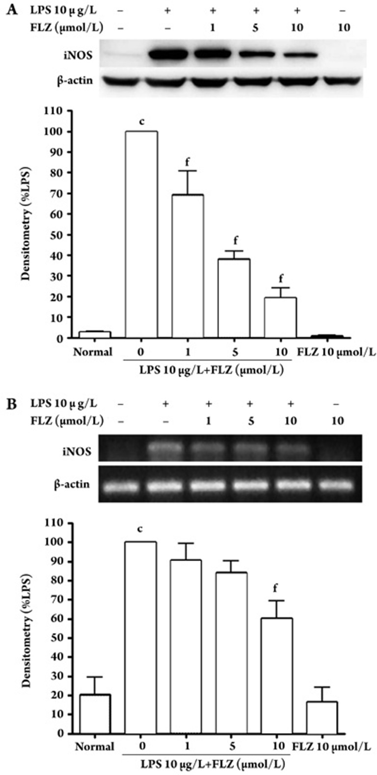
Figure 3.
Effect of FLZ on LPS-induced iNOS protein and mRNA expression in RAW264.7 cells. (A) iNOS protein was detected by Western blot. RAW264.7 cells were pretreated with various concentrations (1, 5, and 10 μmol/L) of FLZ for 30 min; then LPS (10 μg/L) was added and the cells were incubated for 24 h. Total cellular proteins (40 μg) were subjected to Western blot analysis using an antibody against iNOS. (B) RT-PCR detection of iNOS mRNA. RAW264.7 cells were pretreated with different concentrations (1, 5, and 10 μmol/L) of FLZ for 30 min; then LPS (10 μg/L) was added and the cells were incubated for 8 h. iNOS mRNA was detected with RT-PCR and agarose gel electrophoresis as described under Methods. n=3. Values are presented as the mean±SD. cP<0.01 compared with the normal group; fP<0.01 compared with the LPS-treated group.

