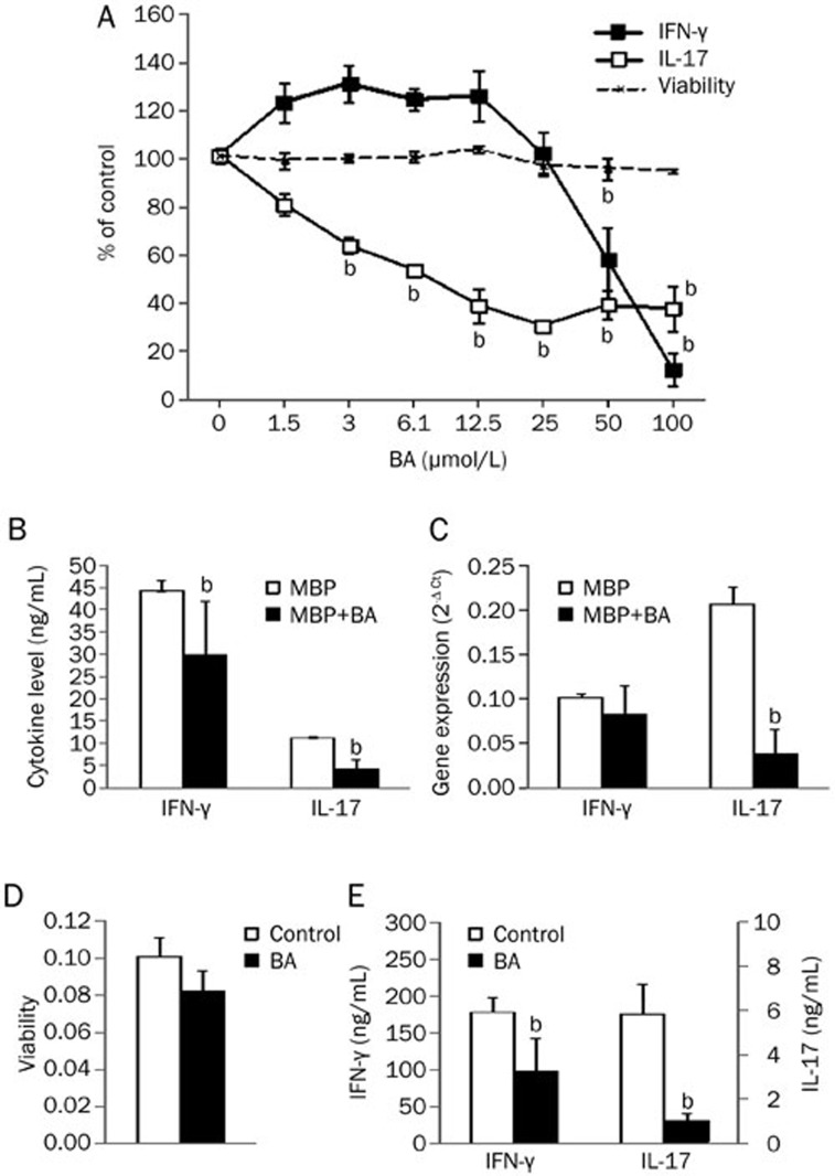
Figure 1.
The influence of BA on IFN-γ and IL-17 generation and viability of MBP-specific T cells. Rats were immunized with MBP and CFA. DLNC were obtained from popliteal lymph nodes at 8–10 d after immunization. SCC were obtained from spinal cords at the peak of EAE. DLNC (2.5× 10⁶/mL) were stimulated with MBP (10 μg/mL) in the presence of various concentrations of BA (A) or 50 μmol/L BA (B, C). SCC were cultivated without stimulation in the absence or presence of 50 μmol/L BA (D, E). After 24 h of cultivation, cell-free culture supernatants were collected for cytokine determination (A, B, E), cell viability was measured by AP assay (A) or MTT assay (D), and DLNC were used for RT-PCR (C). Results are presented as means±SD of values obtained in samples from two (A), eight (B), or three (C, D, E) rats. bP<0.05 represents a statistically significant difference between the cultures grown in the absence (Control) and presence of BA.

