Figure 3.
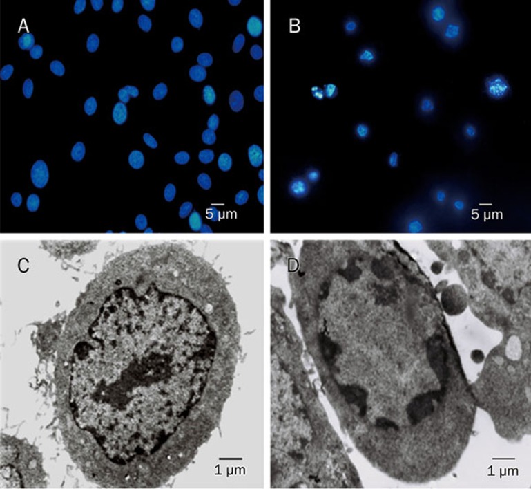
C6 glioma cells displayed apoptotic morphological features after incubation with MG-132. Fluorescence microscopy (A: control group and B: MG-132 group) and transmission electron microscopy (C: control group and D: MG-132 group) showed chromatin condensation, nuclear condensation and nuclear fragmentation in the nucleus of C6 glioma cells treated with MG-132.

