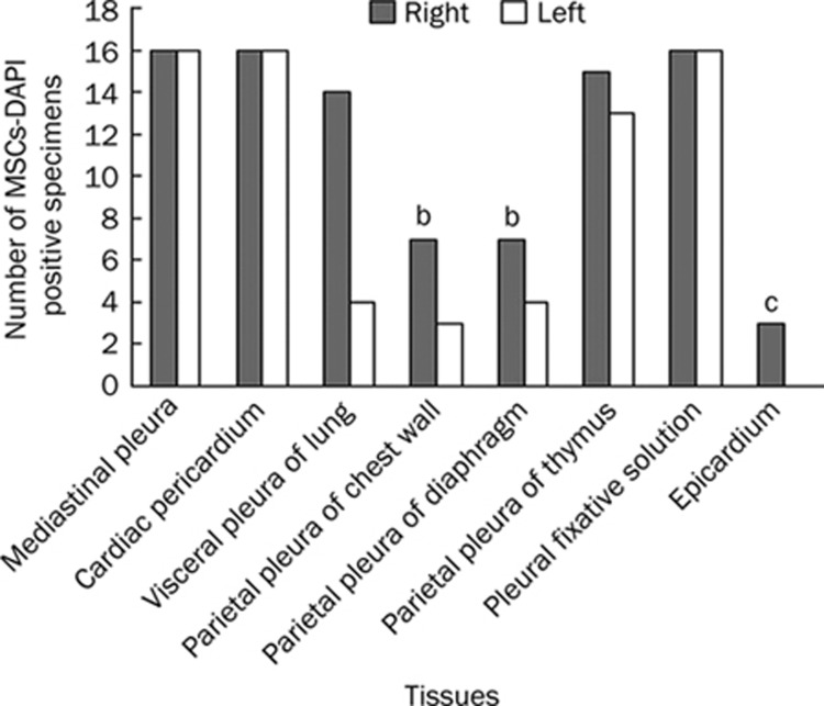
Figure 4.
The MSCs-DAPI positive number of different tissues within two pleural cavities and pleural fixative solutions collected from total 16 experimental rats. For each bar, the maximum number was 16, since the tissue it represents was collected from 16 rats. While DAPI-labeled cells were found in all 64 mediastinal pleura specimens including the cardiac pericardium (100%) and in all 32 pleural fixative solutions (100%), they were found less frequently in other tissues like the epicardium (3/16=19%), the parietal pleura of both left and right chest wall (10/32=31%) and both left and right diaphragm (11/32=34%). In this figure, the column of mediastinal pleura represents only pleura located at both sides of right lung accessory lobe, while pericardium, which also belongs to the mediastinal pleura, was shown in a separate column. Right or Left, specimens collected from the right or left side of pleural cavities. bP<0.05, cP<0.01, compared with the mediastinal pleura including the pericardium.

