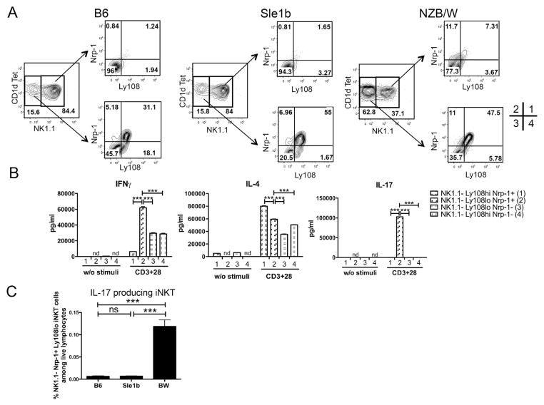
Figure 3.
NK1.1−Ly108loNrp-1+ iNKT cells are IL-17 producing iNKT cells. (A) Ly108 and Nrp-1 expression on gated NK1.1+ and NK1.1− iNKT cells from young B6, Sle1b and BW mice. (B) IFNγ, IL-4 and IL-17 production by sorted BW NK1.1−Ly108hiNrp-1+ (indicated as population 1 as shown in A), NK1.1−Ly108lo Nrp-1+ (population 2), NK1.1−Ly108loNrp-1− (population 3), NK1.1−Ly108hiNrp-1− (population 4) iNKT cells. Cells were sorted from three to five pooled spleens from young BW mice and cultured (5X10⁵/ml) for 3 days with anti-CD3 and CD28 mAbs. (C) Percentages of IL-17 producing iNKT cells, defined as NK1.1−Ly108loNrp-1+TCR+CD1dTet+ population, among live lymphocytes of B6, Sle1b and BW mice. nd, not detected. Group differences with P>0.05 was not considered statistically significant (ns); ***, P<0.001 (two-tailed Student’s t-test). Bar graphs show mean± s.e.m. Results are representative of four independent experiments (A), or two independent experiments (B), or four independent experiments (C). Independent experiments showed similar p values for group comparisons.

