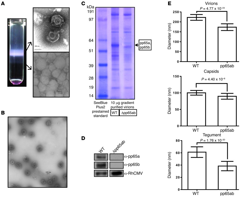
Figure 2. Intact and defective viral particles are secreted from fibroblasts infected with Δpp65ab.
(A) Image of a Nycodenz gradient loaded with RhCMVΔpp65ab, and electron microscope images of virions (top image) and defective particles (bottom image) contained in the visible bands of the gradient. (B) Electron microscope image of purified RhCMVΔpp65ab virions showing the purity of the sample. (C) Purified RhCMV WT and Δpp65ab virions were lysed, and 10 μg protein was electrophoretically separated using NuPAGE MOPS gradient gels and visualized by Coomassie blue staining. (D) Western blots of 5 μg gradient-purified RhCMV 68-1 WT and viral mutant Δpp65ab stained for RhCMV pp65a, pp65b, or a RhCMV-specific antibody. (E) Various electron microscopy images of purified WT and Δpp65ab virions were taken, and the diameters of virions, capsids, and the tegument were determined in multiple images and magnifications (WT, n = 39; Δpp65ab, n = 45). The mean diameters with their respective SDs are shown, and Student’s t tests were performed to determine the P values. Scale bars: 100 nm.

