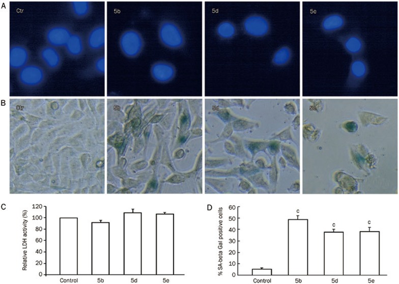
Figure 4.
(A) Detection of apoptosis by Hoechst 33258 staining of A549 cells treated with 5b, 5d, and 5e (80 μmol/L) for 48 h (n=3). Microscopic photographs (×400) were taken under a fluorescence microscope (Nikon). (B) Senescence-associated beta-galactosidase (SA-β-Gal) activity in A549 cells treated with 5b, 5d, and 5e (80 μmol/L) for 48 h. Microscopic photographs (×400) were taken with a phase contrast microscope (Nikon). (C) Effects of compounds 5b, 5d, and 5e (80 μmol/L) on the release of LDH from A549 cells at 48 h (P>0.05 vs control group, n=3). (D) Percentage of SA-β-Gal-positive cells (cP<0.01 vs control group; n=3).

