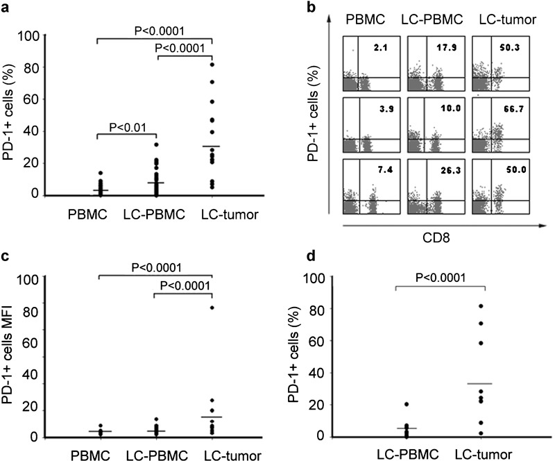
Figure 1.

PD-1 is highly upregulated by tumor-infiltrating CD8+ T cells in patients with NSCLC. (a) Frequencies of PD-1-expressing CD8+ T cells in 58 blood samples, 16 lung tissue samples for LC patients and 23 blood samples for healthy controls. Each dot represents one individual. (b) PD-1 staining representative dot plots for one blood sample from a healthy person, as well as one blood sample and one tumor tissue sample from LC. Values in the upper right quadrant indicate the percentage of cells that express PD-1. (c) MFI of PD-1 expression on CD8+ T cells in PBMC and lung tissue samples for LC patients and healthy controls. Each dot represents one individual. (d) Positive rate of PD-1 in PBMCs and lung tissues from the same individual for 16 LC patients with. LC, lung cancer; MFI, mean fluorescence intensity; PBMC, peripheral blood mononuclear cell; PD-1, programmed death-1.
