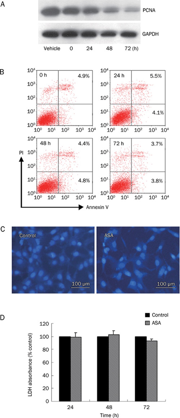
Figure 2.
Effect of ASA on PANC-1 cells proliferation, apoptosis and necrosis. (A) PANC-1 cells were treated with 4 mmol/L ASA for various time and cell lysates were subjected to Western blot using anti-PCNA and anti-GAPDH antibodies. (B) AnnexinV/propidium iodide double staining for apoptosis assay. (C) Fluorescent staining of nuclei by Hoechst 33258 in PANC-1 cells treated with or without 4 mmol/L ASA for 24 h. (D) PANC-1 cells were incubated with 4 mmol/L ASA for the indicated time and cultured medium was collected for LDH release assay. Results represent the mean of three different experiments with quintuple wells.

