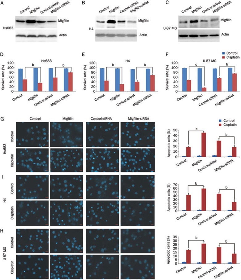
Figure 1.
Regulation of migfilin sensitizes cisplatin-induced apoptosis. (A) Hs683 cells, (B) H4 cells, and (C) U-87 MG cells were examined for expression levels of migfilin by Western blotting after transfection of control vector, pEGFP-C2-migfilin vector, control siRNA, and migfilin siRNA, respectively. (D) Hs683 cells, (E) H4 cells, and (F) U-87 MG cells were examined for the effects of migfilin expression on cell viability and were analyzed by the MTS viability assay. Cells were treated with cisplatin for 24 h. (G) Hs683 cells, (H) H4 cells, and (I) U-87 MG cells were analyzed by DAPI staining for cisplatin-sensitivity. Apoptotic cells were stained with light blue (400×objective). The scale bars stand for 10 μm. Mean±SD. n=3. bP<0.05, cP<0.01 vs control.

