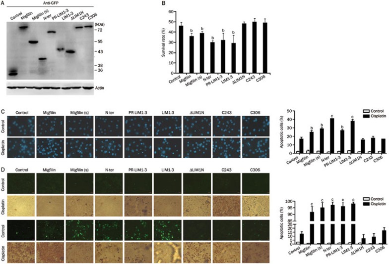
Figure 4.
Functional domains of migfilin were determined in U-87 MG cells. (A) Cellular extracts of transfectants of migfilin mutant vectors with the GFP flag were detected for expression levels of GFP by Western blotting. (B) The MTS viability assay was performed to demonstrate the cell viability of transfectants of migfilin, migfilin(s), N-ter, PR-LIM1-3, LIM1-3, ΔLIM1N, C243, and C306 with cisplatin treatment (n=3. Mean±SD). (C) Apoptotic cell rates of migfilin-mutant transfectants with or without cisplatin treatment were examined by DAPI staining. Apoptotic cells were stained with light blue (400×objective; n=3. Mean±SD). (D) Using the TUNEL staining assay, apoptotic cells of migfilin-mutant transfectants with or without cisplatin treatment were determined by both DAB-stained positive areas (brown areas) and fluorescein-stained areas (green areas) (400×objective). Apoptotic cell rates were calculated under at least 9 different views. Mean±SD. n=3. bP<0.05, cP<0.01 vs control.

