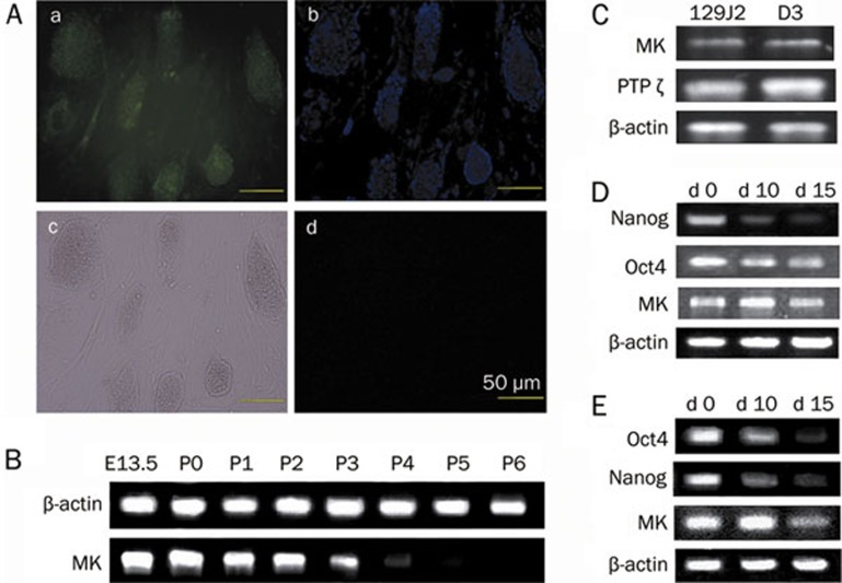
Figure 1.
The presence of midkine (MK) and the MK receptor (PTPζ) in mESCs and MEFs. (A) Immunochemical staining of the mESC line D3 and MEFs with MK antibody. (a) Immunochemical staining. (b) Cell nuclei staining with Hoechst33258. (c) Optical microscopy. (d) Isotype control, scale bars=50 μm. (B) The expression levels of MK in embryos of 13.5 d (E13.5) and MEFs at passage 0–6 (P0–P6). (C) Determination of MK and MK receptor (PTPζ) expression in D3 and 129J2 cell lines by RT-PCR examination. (D) Changes of MK expression during spontaneous differentiation of monolayer cultures. (E) Changes of MK expression during differentiation of EB development. Oct4 and Nanog were stemness markers used to evaluate the differentiation state of mESCs. The housekeeping gene, β-actin, was used as an internal control.

