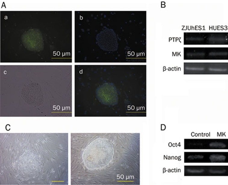
Figure 6.
MK enhances self-renewal of hESCs. (A) The presence of MK protein in hESCs (HUES3): (a) Immunochemical staining. (b) Cell nuclei staining with Hoechst33258. (c) Optical microscopy. (d) Overlap of (1) and (2). (B) The expression of MK and MK receptor PTPζ in hESCs (β-actin as an internal control). (C) Without additional bFGF in hESC medium, 40 ng/mL MK promoted self-renewal of hESCs on feeders (right), control without MK (left). (D) the expression of pluripotency markers Oct4 and Nanog (β-actin as an internal control). Scale bars=50 μm.

