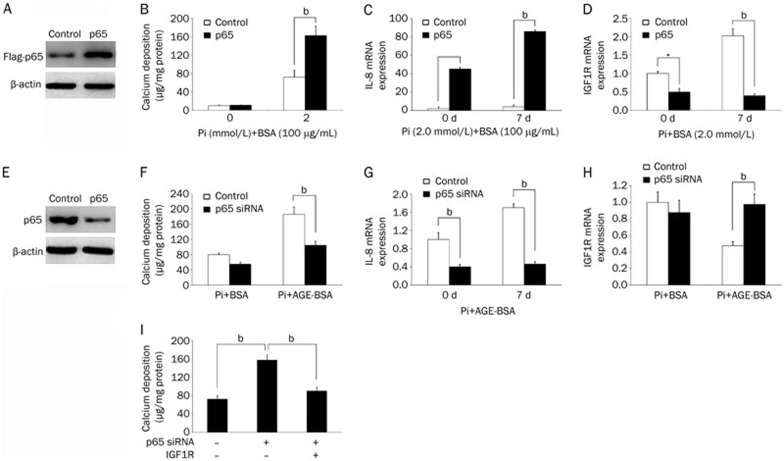
Figure 4.
NF-κB signaling mediates the effects of AGEs on IGF1R expression and calcification in HASMCs. (A) Western blot analysis confirmed the overexpression of Flag-tagged p65 via a lentiviral vector in HASMCs. (B) HASMCs stably expressing p65 or a control empty vector were treated with BSA or AGE-BSA (100 μg/mL) in calcification medium for 7 d (calcium content assay for calcium deposition). (C, D) HASMCs stably expressing p65 or a control vector were treated with BSA or AGE-BSA (100 μg/mL) in calcification medium for 7 d (q-PCR analysis of IL-8 and IGF1R mRNA levels). (E) Western blot analysis confirmed p65 knockdown via a lentiviral vector in HASMCs. (F) HASMCs stably expressing p65 or control siRNA were treated with BSA or AGE-BSA (100 μg/mL) in calcification medium for 7 d (calcium content assay for calcium deposition). (G, H) HASMCs stably expressing p65 or control siRNA were treated with BSA or AGE-BSA (100 μg/mL) in calcification medium for 7 d (q-PCR analysis of IL-8 and IGF1R mRNA levels). (I) HASMCs stably expressing p65 and/or IGF1R via a lentiviral vector were grown in calcification medium for 7 d (calcium content assay for calcium deposition). The data are presented as the mean±SD of three independent experiments performed in duplicate. bP<0.05.

