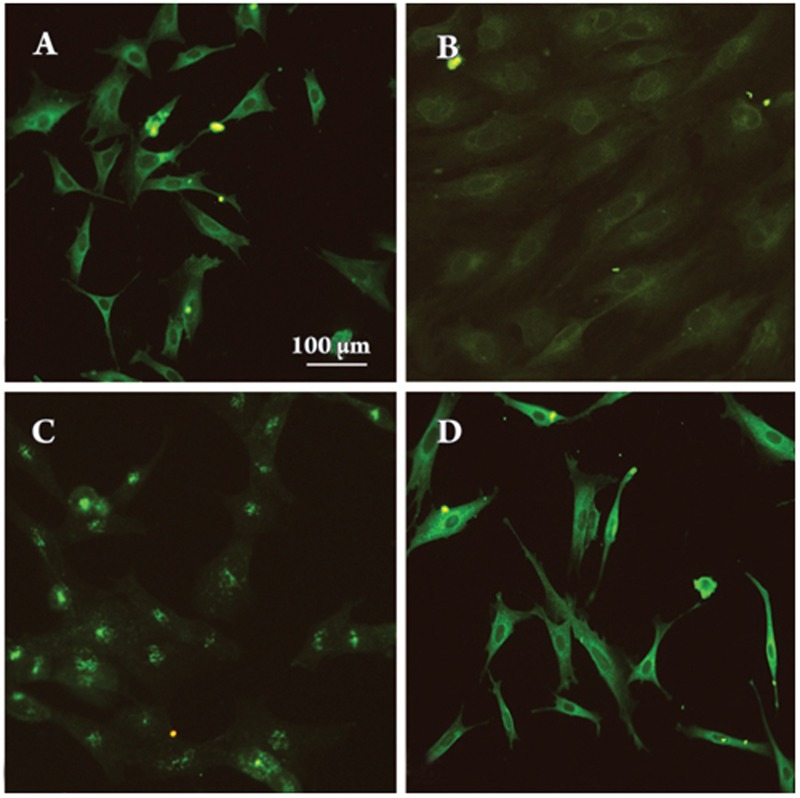
Figure 3.
Subcellular localization of NFATc1. Quiescent VSMCs (A, control) were treated with PE (10 μmol/L) for 9 h (B), 24 h (C) or after pretreatment with PE for 24 h and then incubated with CsA (0.5 μg/mL) for another 24 h (D). All cells at the various times were subjected to immunofluorescence staining using an anti-NFATc1 monoclonal antibody (sc-7294).

