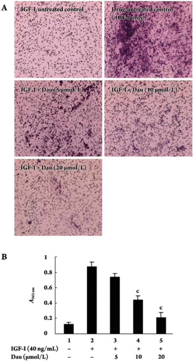
Figure 8.
Effect of Dau on IGF-I-induced invasion of HUVECs in vitro. (A) HUVECs (5×10⁴ cells) were added to the inner chamber of the insert in 300 μL of serum-free media. 500 μL of serum-free media containing IGF-I, at a final concentration of 40 ng/mL, was added to the lower chamber. The cells were cultured for 48 h at 37 °C in the presence or absence of Dau. The invasive cells that migrated from the upper to the lower surface of the membrane were stained and photographed using a computer imaging system. (B) The stained invasive cells were extracted with 200 μL of extract solution, and their absorbance was determined at 562 nm. Data represent mean±SD of three independent experiments. cP<0.01 vs drug-untreated control (IGF-I only, lane 2).

