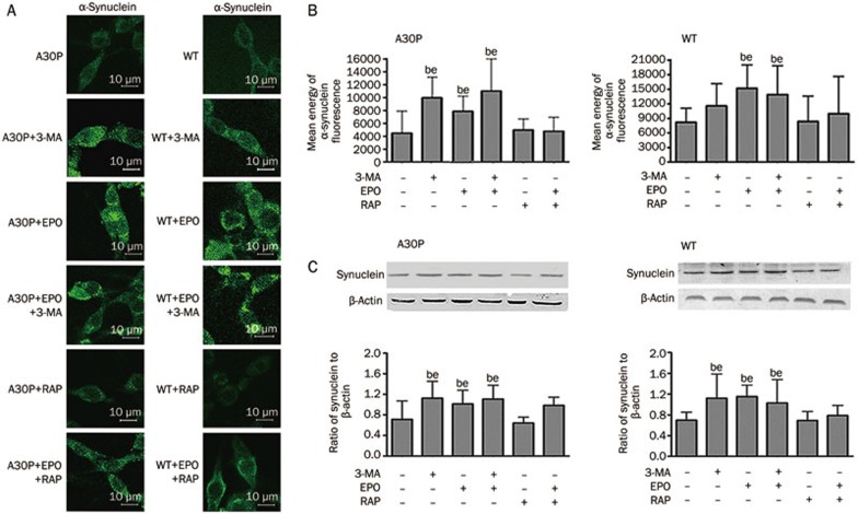
Figure 2.
(A) Cells were imaged by confocal microscopy. A30P and wild-type cells treated with the indicated drugs were permeabilized and immunostained with anti-α-synuclein followed by FITC-labeled secondary antibodies for imaging. (B) The immunofluorescence mean energy of cells was analyzed. At least 50 cells in each group were randomly selected using the same settings. The data were normally distributed and statistically analyzed using one-way ANOVA. (C) Western blot analysis of total exogenous human α-synuclein in A30P and wild-type PC12 cell lines. Mean±SD. bP<0.05 vs control. eP<0.05 vs RAP.

