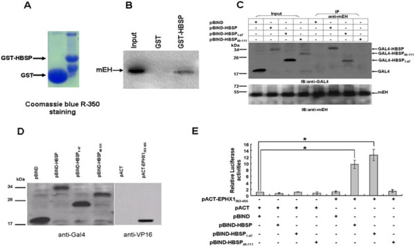
Figure 1.

In vitro and In vivo interaction between HBSP and mEH. (A) Bacterially expressed GST and GST-HBSP recombinant proteins bound to glutathione-sepharose beads. (B) GST pull-down assay. GST recombinant proteins were incubated with 35S-labeled mEH, immobilized proteins and subjected to SDS-PAGE and autoradiography. (C) Co-IP assay showing the interaction between HBSP and endogenous mEH in Huh-7 hepatoma cells. Cells were separately transfected with pBIND-HBSP, pBIND-HBSP1–47, or pBIND-HBSP48–111. Cell lysates from transfected cells were immunoprecipitated with anti-mEH, and the immunoprecipitation complexes were subjected to immunoblotting with anti-GAL4BD (upper) or anti-mEH (lower). (D) Western blot analysis of full-length and truncated HBSP, expressed as a fusion protein, with GAL4 DNA binding domain (left). mEH353-455 expressed as a fusion protein, with VP16 activation domain in Huh-7 hepatoma cells (right). (E) Mammalian two-hybrid assay. HuH-7 cells were lysed 48 h after transfection and renilla-normalized firefly luciferase activity was determined using the dual luciferase assay system. Data are presented as means ± SD for three independent experiments. The firefly luciferase expression is given as folds over the background (set arbitrarily at 1). (* P < 0.01).
