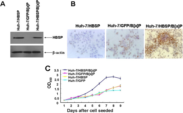
Figure 3.

Effects of HBSP on the proliferation of B[alpha]P-treated Huh-7 hepatoma cells. (A) HBSP expression in Huh-7/HBSP/B[alpha]P, Huh-7/GFP/B[alpha]P or Huh-7/HBSP. 30 μg of cellular proteins were subjected to 12% SDS-PAGE, transfered to a PVDF membrane, and probed with anti-mEH. β-actin served as a loading control. (B) Detection of BPDE-DNA in Huh-7/HBSP/B[alpha]P, Huh-7/GFP/B[alpha]P or Huh-7/HBSP cells. Cells on sterilized glass coverslips were detected for BPDE-DNA by immunocytochemistry assay. Images were taken at × 400 magnification. (C) Cell proliferation of Huh-7/HBSP/B[alpha]P, Huh-7/GFP/B[alpha]P, Huh-7/HBSP or Huh-7/GFP cells. Cells were seeded in 96-well plates at 2 × 10³/well, cell proliferation was determined daily in triplicate for 9 days by BrdU assay. The optical density (OD) was measured at 450 nm using a microplate reader. The analyses were repeated three times, and the results were expressed as mean ± SD.
