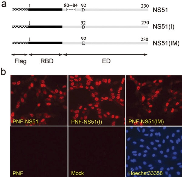
Figure 1.

Three H5N1-NS1 variants and their subcellular localization. (a) Schematic diagram of H5N1-NS1 variants. The mosaic bars and Flag represent the Flag tag at the N terminus of NS1. The solid black bars and RBD indicate the RNA-binding domain mapped between residues 1 and 73. The solid gray bars and ED indicate the effector domain at 74–230 residues. NS51: H5N1-NS1 variant with a 5-aa deletion at position 80–84; NS51(I): H5N1-NS1 variant with a 5-aa insertion at the corresponding position; NS51(IM): H5N1-NS1 variant carrying both the 5-aa insertion and a D92E mutation. (b) Intracellular distribution of H5N1-NS1 variants in Hela cells. Cells were transfected with indicated plasmids or empty PNF vector. At 24 h post-transfection, cells were fixed, permeabilized and stained with rabbit anti-Flag antibody and Cy3-conjugated goat anti-rabbit secondary antibody followed by nuclear staining with Hoechst 33258. aa, amino acid; NS1, non-structural protein 1.
