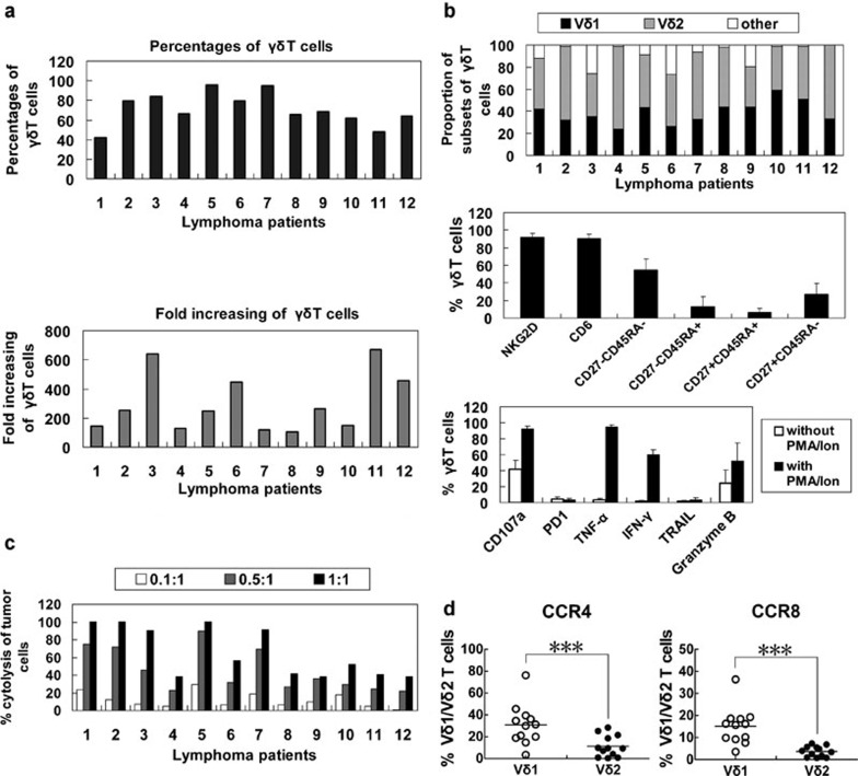
Figure 5.
Proliferation, phenotype and cytotoxicity of γδ T cells from the peripheral blood of lymphoma patients. (a) The purity and proliferation efficiency of in vitro expanded γδ T cells of lymphoma patients (n=12). (b) Phenotype expression and cytokine release by γδ T cells (n=12). (c) Cytotoxicity of γδ T cells from 12 lymphoma patients. (d) Percentages of CCR4+ and CCR8+ cells among the Vδ1 and Vδ2T cells from patients. ***P<0.005. CCR, C–C chemokine receptor; IFN, interferon; PD1, programmed-death receptor 1; TNF, tumour-necrosis factor; TRAIL, TNF-related apoptosis-inducing ligand.

