Figure 1.
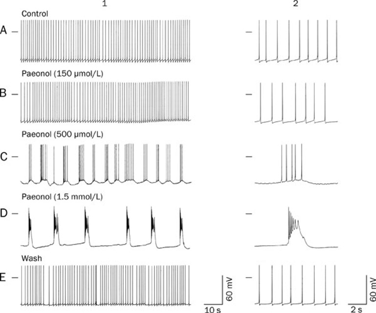
Effects of paeonol on the RP4 neuron. A, B, C, D, and E were recorded from the same neuron. A1: Control; the neuron shows spontaneous firing of action potentials. B1, C1, and D1: Membrane potentials recorded 20 min after the administration of paeonol (150, 500 and 1500 μmol/L), respectively. E1: Membrane potentials measured at 30 min after the washing off of paeonol from D1. A2, B2, C2, D2, and E2: Expanded pictures showing individual action potentials related to A1, B1, C1, D1, and E1, respectively. The horizontal bar on the upper left side indicates the membrane potential at 0 mV. Notably, action potential bursts were not elicited by paeonol at 150 μmol/L but were reversibly elicited at concentrations of 500 μmol/L and 1.5 mmol/L.

