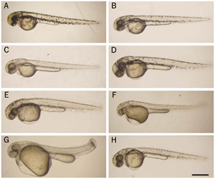
Figure 1.
Morphological changes induced by dihydroartemisinin (DHA) and artemisinin during zebrafish early embryonic development. Embryos treated with different concentrations of compounds were imaged at 48 hpf. (A) No treatment; (B) 0.1% DMSO; (C) 1.0 mg/L DHA; (D) 2.5 mg/L DHA; (E) 5.0 mg/L DHA; (F) 10 mg/L DHA; (G) 15 mg/L DHA; (H) 10 mg/L artemisinin. The scale bar represents 500 μm for all panels.

