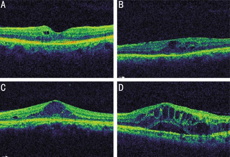
Figure 1. DME classification based on OCT appearance.
A: Patients with focal DME had limited retinal thickening with cyst formation and preservation of the macular contours; B: Diffuse DME patients had widespread retinal thickening with a sponge like appearance of the macula; C: Patients with focal cystoid DME had a mound like appearance of the fovea due to focal collection of fluid at the fovea; D: Patients with neurosensory detachment DME had an associated subretinal collection of fluid under the fovea.

