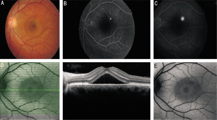
Figure 1. Images of a case with acute CSCR.
A: Color fundus photography of an acute CSCR 37-year-old male case shows serous detachment; B,C: FA of the same case shows hyper-fluorescence at the leakage site which begins at the 35th second and increases in late phases; D: Horizontal section of OCT of the same case shows serous detachment with distinct border but without any changes in the outer segments of photoreceptors and serous fluid; E: Fundus autofluorescence photography reveals hypo-autofluorescence in the serous detachment area with distinct borders and also hypo-autofluorescence at the leakage site in FA surrounded by iso-autofluorescence area of normal retina.

