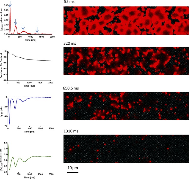
Figure 11.
Behavior of the calcium clock in isolation. The model cell was voltage clamped to −50 mV starting from a state with 1 mM of free calcium in the SR. The decaying oscillations actually consist of LCR events temporarily synchronized by the initial state. With time, LCR events become less synchronous, and cell calcium declines as a result of extrusion of calcium by NCX. The eventual steady state still consists of high calcium local events propagated over limited distances.

