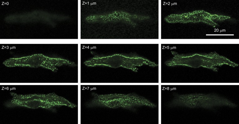
Figure 7.
Serial confocal sections of a SANC stained with fluorescent antibody to RyR. RyRs are limited to the surface membrane (this pattern was observed in 15 out of 25 cells). Tangential sections show that surface RyRs form an irregular network of large and small RyR clusters. Confocal sectioning was performed (along the z axis) with a step of 1 µm (Z = 0 at the bottom).

