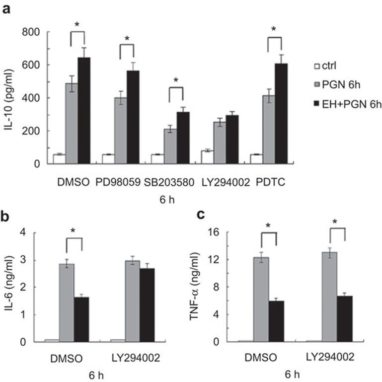
Figure 5.
Blockade of PI3K abrogates EH-induced IL-10 expression and decreased IL-6 expression in macrophages. (a) Mouse peritoneal macrophages (3×10⁵ cells/300 µl) were seeded the day before stimulation. Cells were pretreated for 30 min with DMSO, ERK inhibitor PD98059 (10 µM), p38 inhibitor SB203580 (10 µM), PI3K inhibitor LY294002 (20 µM) or NF-κB inhibitor PDTC (20 µM), and then stimulated with PGN (25 µg/ml) and EH (7.5 µg/ml) as indicated for 6 h. IL-10 was measured in the supernatants by ELISA. The data are shown as the mean±s.d. of three independent experiments. * indicates P<0.05. (b) Cells were pre-treated for 30 min with DMSO, PI3K inhibitor LY294002 (20 µM), and then stimulated as indicated for 6 h. IL-6 (B) and TNF-α (c) levels in the supernatants were measured by ELISA. The data are shown as the mean±s.d. of three independent experiments. * indicates P<0.05. EH, ephedrine hydrochloride; ERK, extracellular signal-regulated kinase; PGN, peptidoglycan; PI3K, phosphatidylinositol 3-kinase; NF-κB, nuclear factor-κb; s.d., standard deviation; TNF, tumor-necrosis factor.

